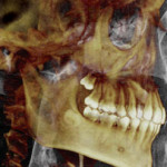
Cone Beam CT
Cone Beam Computed Tomography | CBCT
 Cone beam computed tomography is one of the cutting edge diagnostics that are available at Oral and Maxillofacial Surgeons of Houston. CBCT is a medical imaging technique of producing a three-dimensional image of the internal structures of a solid object (such as the jaw and teeth). The X-rays are divergent, or opposing, and form a cone. For the assessment and planning of surgical implants, a dental cone beam scan can offer vital information for the surgeon to consider.
Cone beam technology has been used since 1998. Its advancement has warranted it to become an increasingly important component of the diagnosis and treatment planning of the implant dentistry we do at Oral and Maxillofacial Surgeons of Houston.
The 3D image that can be generated by this cone beam technology presents an undistorted view of the development of teeth (dentition). This image can be used to accurately visualize:
- erupted teeth
- non-erupted teeth
- tooth root orientation
- irregular structures that conventional 2D radiography cannot
The American Academy of Oral and Maxillofacial Radiology (AAOMR) now suggests cone-beam CT to be the preferred method for pre-surgical assessment of dental implant sites, and we already utilizing this technology. The professionals at Oral and Maxillofacial Surgeons of Houston are highly trained experts of CBCT.
